
Overview of Services
|
|

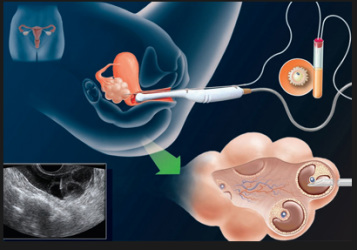
PGD: Pre-implantation Genetic Diagnosis:
embryo biopsy is the more practical method for pre-implantation genetic diagnosis. The biopsy can be performed at one of the following stages:
Cleavage-stage Embryos This is usually performed on day 3 embryos (i.e. 3 days after oocyte retrieval) which have normally reached the 6 to 10 cell stage. Under the inverted microscope used for micromanipulation the embryo is held by gentle suction from one side using the holding pipette. On the opposite side of the embryo, a hole is made in the zona pellucida either mechanically using a fine-glass knife or by applying acid Tyrode’s solution locally using a fine pipette. A fine-bore glass pipette can then be pushed through the hole and one or two blastomeres aspirated.
Blastocyst Stage Embryos Here the biopsy is taken on day 5 or 6 (i.e. 5 or 6 days after oocyte retrieval). At this stage, the embryo has reached the blastocyst stage. The advantages of this technique is that more cells may be used for biopsy. In this technique, 3 to 5 cells are usually aspirated after drilling a hole in the zona pellucida using acid Tyrode’s solution. These cells are taken from the trophectoderm at the anembryonic pole away from the inner cell mass which will develop into the fetus.
|

Laparoscopic Surgical Evaluation and Treatment
|